Animal cell
Part1
Structure and function
 |
| Simple figure of eukaryotic cell |
Introduction
Cellular Level of Organization
The Cell Theory
In the 1830s, Matthias Schleiden studying plants and Theodor Schwann studying animals independently declared these organisms were made of cells. All organisms, both unicellular and multicellular, are made up of cells. cells are the smallest units of living matter and are the structural and functional units of all organisms. Rudolf Virchow declared cells come only from pre-existing cells. So, Cells are capable of self-reproduction.
Cell size, Cells range in size from a frog's egg ( one millimeter) down to one micrometer. Cells need a surface area of plasma membrane large enough to adequately exchange materials.
Note: Largest human cells are the Ovum ( Egg cells) about 120 micrometers, skin cells 30 - 35 micrometers, red blood cells about 5 micrometers and sperm cells which are considered to be the smallest ranges from 4 to 4.5 micrometers
Surface-area-to-volume ratio......dictates that cells must be small
As cells get larger in volume, surface area relative to volume decreases. Size limits how large the actively metabolizing cells can become. Cells that need greater surface area use modifications such as folding and microvilli.e.g, Macrophages ( a type of our innate immune cells that functions as a cell-eater, it can devour many bacterial cells). We have 2 major types of cells, Prokaryotic and Eukaryotic
Prokaryotic cells
All prokaryotic cells lack a nucleus and are smaller and simpler than eukaryotic cells. Prokaryotic cells were the first cells and date back to earliest evolutionary history; Because they are biochemically different, prokaryotes are divided into 2 domains: Bacteria and Archaea
Eukaryotic cells
Eukaryotic cells are members of the domain Eukarya, including kingdoms Fungi, Animalia, Plantae, and Protista. A membrane-bounded nucleus houses DNA. The nucleus may have originated as an invagination of the plasma membrane.
The structure of our cells
-Organelle.., word named on organs with a considerable small size which is why it is mainly introduced to cell organs, they are membrane-bounded organelles ( consisting of phospho-bilipid layer) just like the own membrane of the cell, this membrane separates the organelle from the area around it inside the cell.
-A single cell contains many organelles that provide various and crucial functions to our body, the group of cells comprise tissue, a group of tissues makes up an organ, many different organs builds up a body so we conclude that cells are our building units but what is inside this cell?
- To begin with, a cell contain lysosomes, mitochondria.p,(mitochondrion.singular,), vacuoles, nucleus, endoplasmic reticulum ( smooth & rough ), Golgi apparatus, ribosomes, centrioles, cell membrane and last but not least cytoplasm)
2) Nucleus, the headquarters of a cell, the mastermind of a cell surrounded by a nuclear membrane, a densely packaged chromatin found inside of it ( our genetic material) which can be unfolded reaching up to 2 meters, chromatin ( is the double stranded DNA + histone proteins which is responsible for the dense packaging to fit inside) the nucleus average diameter is 6 micrometers, nucleolus is a part of the nucleus through which the ribosomes are manufactured. (Pic.source:https://epiehonorsbiology.wikispaces.com/Nucleus2?responseToken=0d241e85e112dc01d3c1c27fa3ee7cfdc)
3) Mitochondrion, spherical body, with outer and inner membranes ( intermembrane space found between them ) folded from the inside, area inside the folds is called Matrix while the outside is the crista, has ribosomes and its own DNA ( it is an independent organelle ) 0.5 to 2 microns in length (Pic.source: http://faculty.une.edu/com/abell/histo/histolab2.htm)
4) Lysosomes, membrane-bounded organelle, small-rounded bubbles containing essential enzymes manufactured by the endoplasmic reticulum. (Pic.source: http://quatr.us/biology/cells/lysosome.htm )
3) Mitochondrion, spherical body, with outer and inner membranes ( intermembrane space found between them ) folded from the inside, area inside the folds is called Matrix while the outside is the crista, has ribosomes and its own DNA ( it is an independent organelle ) 0.5 to 2 microns in length (Pic.source: http://faculty.une.edu/com/abell/histo/histolab2.htm)
 |
| Mitochondrion simple anatomy - made by me |
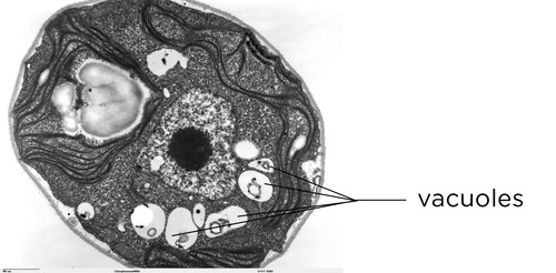
5)Vacuole, a space or vesicle within the cytoplasm of a cell, enclosed by a membrane and typically containing fluid. (Pic.source: http://www.mstworkbooks.co.za/natural-sciences/gr9/gr9-ll-01.html )
6)Endoplasmic reticulum ( rough and smooth ), mainly a network of flattened minute tubular or irregular in shape and branches and are surrounded by thin membranes found in the cytoplasm forming many folds, RER ( rough endoplasmic reticulum) named rough due to the attachment of ribosomal units on its membranes, SER ( smooth endoplasmic reticulum) named smooth due to the absence of ribosomal units, the ribosomes appear like dots on the surface of the endoplasmic reticulum ( rough ) - it is the second organelle after nucleus in its volume as it takes over a remarkable volume in the cytoplasm )
(Pic.source: https://www.uni-mainz.de/FB/Medizin/Anatomie/workshop/EM/EMSERE.html
http://www.cellimagelibrary.org/images/37237)
7) Ribosomes, are small rounded circular units mainly built of RNAs, spread all over the cell in many numbers and are found on endoplasmic reticulum giving it its rough structure, has many types tRNA, rRNA and mRNA. (Pic.source: https://www.uni-mainz.de/FB/Medizin/Anatomie/workshop/EM/EMVokabular/EMR.html )
8) Centrioles, cylindrical/rod-like structures found nearby the nucleus of animal cells, they perform major functions during cell division - they have the ability to produce thin fibres known as spindle fibers, its outer wall contains a number of microtubules arranged in 9 groups each of them is formed of 3 microtubules - 0.2 to 0.8 micrometre in length and 0.2 micrometres in diameter.
(Pic.source: http://www.biology-pages.info/C/Centrioles.html )
9)Cytoplasm, It is the swimming pool for all the previous organelles, it is where the organelles are kept inside, proteins to be vesicled are found, ribosomes are distributed everywhere - jelly-like substance, a water-based solution containing that contains ions, small molecules, and macromolecules-
(Pic.source: https://www.uni-mainz.de/FB/Medizin/Anatomie/workshop/EM/EMCytoplasmaE.html )
10) Cell membrane, Phospho-bilipid layer, consists of hydrophilic head ( hydro= water, philic= loving or attracted ) and hydrophobic tail ( means water-repellent), our cell membrane has a lot of contributions to our body, it functions in homeostasis, transporting substances due to its semi-permeability and being the main reason of cell signaling due to the presence of membrane-bound receptors on the surface, glycoproteins (receptor function), integral and peripheral proteins
Functions
-The cell, which is generally the building unit of our body, its itself being the building unit makes a major function since it has many types, connective tissue forming cells, muscular-forming tissue cells and as well as all of our organs with a few differences, the true cell ( eukaryotic) contains as we know its organelles, each of which has its own function and together all working properly makes up a powerful bio-machine ( cell ) a cell contains more than 50 thousand different proteins essential for its vitality and viability throughout its entire life.-A group of cells together forms a tissue, Moreover, a group of tissues makes up organs, Last but not least, different organs constructs the human body; so as a conclusion, cells are the basic building units but what really makes it the basic structural unit of the human body.
-Basically, from the previous point we discussed the structures of a cell and known that it has organelles, these organelles are the main reason we keep saying that a cell is the building unit of the human body due to the following vital functions carried out by these organelles:
- Mitochondria, it is the powerhouse of the cell as they contain respiratory enzymes, present in both animal and plant cells, energy-rich molecules known as ATP ( adenosine tri-phosphate) are formed and stored in reduced forms known as NADH2 (Nicotinamide adenine dinucleotides) for further use, this organelle has its own DNA meaning it is independent of the cell's nucleus, it doesn't have to follow division if the cell is carrying division, the production of ATPs is carried out by a process known as Krebs cycle (named after the scientist who discovered it Hans Krebs) a.k.a tri-carboxylic acid cycle a.k.a citric acid cycle, Furthermore, this cycle is carried out aerobically ( In presence of oxygen ), Last but not least, such process produce 2 molecules of ATP, 8 molecules of NADH2 and two molecules of FADH2 ( flavine adenine dinucleotide )
- Nucleus, it is the most important structure in a cell, it contains the genetic material ( DNA ) which encodes all the essential and required proteins for the cell to thrive and survive, In the nucleus a chromatin reticulum which is the DNA + histone proteins bound together, it condenses right before the initiation of mitosis in case of cell division to nucleosomes scale level in order to launch the division, also it is where transcription occurs ( a process of which the cell's own DNA acts as a template in making Messenger RNAs ( mRNAs) which is supposed to be the cell needs, inside of this nucleus a large circular portion called nucleolus which is where ribosomal RNAs are synthesized, If our body is in need of a specific combination of proteins/amino acids then such organelle (nucleus) is the one responsible for this job, Besides, the DNA ( genetic material) holds all our phenotypic and genotypic characteristics ( everything) whether if some are in active or inactive form.
- Golgi- apparatus, it receives anything whether synthesized from nucleus, in cytoplasm and is needed to be expelled out of the cell, from the endoplasmic reticulum, it packages it in vesicles (rounded structures) so they can be soon sent to the outside inside the vesicles or disintegrated and released into the blood in some cases.
- Centrioles, these small structures has a great role during cell division, with which it forms the spindle fibers as we know them which binds with chromosome during the phases of cell division and starts pulling them to provide copies of cells having the same and equal genetic material in case of Mitotic cell division or reduction of this genetic material in case of Meiotic division.
- Ribosomes, they have various types mRNA, rRNA and tRNA types of ribosomes, each of which has its own function, all ribosomes contribute in DNA transcription ( which is a process of changing DNA into RNA or to be more clear mRNA ( messenger RNA) and translation which is, in other words, mean ''protein synthesis'' production of amino acids connected to each other by peptide bonds forming a polypeptide chain (proteins), rRNA ( ribosomal RNA) these are units of ribosomes main job is to read the mRNA and provide message for the tRNA ( transfer RNA) to perform its function which is getting the specific amino acid (anti-codon) for the specific (codon - the name of the bases in mRNA) **There will be a whole topic covering all what you need to know about Ribosomes since they have a lot of information**
- Lysosomes, are the digesting units of an animal cell, containing degrading enzymes enclosed in membranes, such organelles work on breaking down any cellular wastes, renew materials and also they are found more in immune function cells contributes in killing any intruders ( e.g. bacteria) by releasing its enzymes on these foreign bodies, They are more abundant in animal cells ( e.g. cells of liver, kidney and small intestines) than plant cells
- Plasma (Cell) membrane, Every cell is surrounded by a membrane which protects its constituents and regulates the relationship between the cell and its surroundings. It also regulates the passage of substances into the cell and out of the cell - it described as a mosaic fluid, due to its selective permeability it regulates the passage of the nutrient materials, thus enabling the cell to choose what it needs from different materials, these substances move through the plasma membrane by diffusion, active transport and phagocytosis - contributes to homeostasis (internal temperature of the body which is normally 37*C) and regulation of electrolytes in the body - cell signaling due to the presence of receptors such as glycoproteins which play an important role in the communication between cells such as antigen-presenting cells to immune cells in order to identify the target vector, or the secretion of any type of substances in order to alert other cells of presence of foreign invaders such as Cytokines produced by Helper T cells in the field of invasion. **We will have a whole topic about Cell signaling since it is so interesting topic**
 |
| Fluid mosaic model of the cell membrane source: http://www.macroevolution.net/fluid-mosaic-model.html |
- Endoplasmic reticulum, It can be found adhered to the plasma membrane of the cell, It may also extend between the cells in order to communicate with the membranes if the neighboring cells. In this way, all the cells are connected to each other through their endoplasmic reticula. In addition to it plays a part in the formation of secretions in the cells, also the ribosomes on its surface are responsible for protein synthesis. Moreover, it acts as an internal transport system that connects together the various parts of the cytoplasm with the nucleus and also with those of other cells.
- Cytoplasm, It functions like the bedding material, the fluid which holds all the cell organelles together also contributing to the shape of the cell, extending from the nucleus to the plasma membrane in eukaryotes, due to its cytoskeleton ''a.k.a cytosol'' a network of fibers about the whole cell giving it shape also organize these cellular components. Elements making this cytoskeleton include actin filaments, intermediate filaments, and microtubules.
- Actin filaments are long, thin fibers ( about 7 nanometres in diameter) that occur in bundles or meshlike networks. Actin filaments move by interacting with myosin: myosin combines with and splits ATP, binding to actin and changing configuration to pull actin filaments forward.
- Intermediate filaments are 8-11 nanometers in diameter, between actin filaments and microtubules in size. They are rope-like assemblies of fibrous polypeptides. Some support the nuclear envelope; others support plasma membrane and form cell-to-cell junctions.
- Microtubules are small hollow cylinders (25 nanometres in diameter and form). and are composed of a globular protein tubulin.
- Cell Vacuole, It helps in storing the wastes resulted from metabolic reactions or any cell activity and removing them in order to protect the cell from any damage due to the accumulation of wastes.
Thanks everyone, I hope you find this informative and if you do please subscribe, comment any questions if you have or perhaps if you would like to share your opinion with me either by writing in comments or voting the poll you will find below every topic I do, don't forget to share it with every biologist :) it really help me out and cheer me up.
#WeliveWelearn
#Biology
Check out my latest topic at -----> http://allaboutbiologyworld.blogspot.com/2017/06/pituitary-gland.html
For more biology stuff----> allaboutbiologyworld.blogspot.com
Facebook group -----> https://www.facebook.com/biologyclub17/
Principles of Zoology course no. 11101
Botany - a classification to Plant kingdom course no. 07102
https://micro.magnet.fsu.edu/index.html
https://www.khanacademy.org/
My highschool biology courses
William Stillwell-An Introduction to Biological Membranes. Composition, Structure and Function-Elsevier Science (2016)
No comments:
Post a Comment