Chatbox

Monday, 29 January 2018

Thyroid gland #Part2 Hormones Physiology & Hyperthyroidism

Thyroid gland

Part 2

Physiology and Grave's disease (hyperthyroidism) 


How does the thyroid gland synthesizes the thyroid hormones?


The hypothalamic pituitary thyroid axis
  First of all, the thyroid gland hormones are T3 (Triiodothyronine), T4 (Thyroxine), and Calcitonin, iodine is an essential element for the synthesis of these hormones, the thyroid gland is situated in front of the trachea below the hyoid bone ( the only floating bone), Moreover, the thyroid gland is very crucial to our body's metabolism at which it tends to make sure that the cells in your body are working properly by instructing every cell in the body when to consume oxygen and nutrients making it responsible for our growth and providence of better health, Furthermore, the calcitonin secreted by the thyroid gland, is an essential hormone that reduces your body's calcium level in the blood when it goes haywire, Ultimately, the thyroid gland allows our cells to use energy, grow and reproduce.

  It begins when a part of the brain called hypothalamus secretes TRH (thyrotropin-releasing hormone) stimulates the pituitary gland to release TSH (thyroid stimulating hormone) which induces the thyroid gland to release its hormones, usually there is a negative feedback when the thyroid hormones are too much in the blood that should eventually inhibit the release of TRH from hypothalamus.

  Now, the thyroid gland consists of lobules that each containing smaller cells called follicles ( follicular cells ) the lumen of these surrounded follicular cells are called colloid. Deep into a cellular level We have the cell membrane of the follicular cells which is surrounded by blood vessels that feed it with oxygen and the needed nutrients, the TSH molecules coming from the pituitary gland through the bloodstream binds to G-protein coupled receptors (GPCRs) present on the cells' membranes which then exchanges GTP to GDP thereby increases the cellular cyclicAMP (adenosine monophosphate) levels essentially increases the production of thyroid hormones ( T3 & T4) that eventually maintains the body's metabolism.

 GPCRs are family of receptors that are found almost in every eukaryotic cell and has various bodily functions in our physiology. ***There will be a specific topic about GPCRs since they have a very important bodily function also it is a heavily researched topic***
But for now, you can understand their mechanism through here --> https://www.youtube.com/watch?v=ZBSo_GFN3qI&t=183s 

How does that happen? The Physiology of Thyroid's hormones

 A quick recap of the previous paragraphs, On a cellular level We have the thyroid gland's follicular cells forming the colloid surrounded by blood vessels which contain all the nutrients and essential elements required for the synthesis of the thyroid's hormones, it contains various cations and anions such as Na+, K+, Ca+2, Cl-, (HCO3)- and I- among other things.

 Inside the follicular cells, the endoplasmic reticulum through which proteins are synthesized, Special proteins are synthesized inside which makes up most of the thyroid gland's proteins '' Thyroglobulin'' dimeric protein, It then travels to the Golgi apparatus to be packaged, After that it is transported to the colloid.
 The follicular cells contain the following NIS '' Sodium and Iodine symporter'' (transporter), sodium and potassium pump and transporter called ''Pendrin''.
The NIS '' Sodium and Iodine symporter'', it allows iodine and sodium ions to go inside the follicular cells from the blood vessels at the same time, simultaneously the sodium can go out when the potassium ions come in; because the thyroid hormones synthesis occurs in the colloid and not in the follicular cells so the Iodide ions need to be transported to the colloid.
Pendrin ''transporter'' it transports the iodide ions to the colloid from the cells which is exchanged by chloride ions at the same time. Moreover, there are Peroxidases that oxidizes the iodide ions to iodine which is, later on, attaches to the Thyroglobulin ( several chains of Tyrosine molecules)

When one iodine molecule attach to thyroglobulin, We have MonoIodoTyrosine   
            Two iodine molecule attach to thyroglobulin, We have DiIodoTyrosine

 If MonoIodoTyrosine (MIT) binds with DiIodoTyrosine (DIT) by ester bonds, we have triiodothyronine. But, If DiIodoTyrosine (DIT) binds with DiIodoTyrosine (DIT), we have Thyroxine, Yet the hormones aren't ready yet since the complex is still a part of the thyroglobulin.
The synthesized complex before will move back to the follicular cells in vesicles via pinocytosis, in which it will be attached to the lysosomes found in the follicular cells, they will bind to the endosome and break down the 2 hormones from the thyroglobulin to be released in the blood.

(Note: They also can be de-ionized into Iodide molecules and Tyrosine)
 Inside the bloodstream, there are thyroid-binding proteins which act as emulsifiers to bind the thyroid hormones ( which are lipid-structures ) that are water-insoluble will bind with the thyroid-binding proteins to be delivered to the certain cells for metabolism regulation.

 Let's take skeletal muscle cells as an example. The cells' membranes composition is lipid-bi-layered so the thyroid hormones goes easily through it, they enter the nucleus and bind to thyroid hormone receptors and retinoid X receptors, this will initiate gene transcription for specific mRNAs that will promote the thyroid hormone response resulting in the release of mRNA to the cytoplasm to be translate as a promotion for the thyroid hormone response ( this is like the feedback of the reception of thyroid hormones to the cells) and Ultimately, increasing the metabolic rate depending on which the area these hormones were attached to.
(Note: T3 is more active than T4, therefore when these hormones reach their destined cells, T4 is converted to T3)

 The thyroid gland has a great impact on us regulating and maintaining our metabolism every bit of second, it promotes the growth of tissues, regulate your heart pumps, maintaining blood pressure, body temperature, triggers the production of digestive juices, and basically the thyroid gland's hormones are mainly concerned with the main thing that a human body cannot do without it which is our Homeostasis. 

 So what would happen if a defect occurred within the thyroid gland that made it secrete its hormones continuously with no inhibition of secretion....

 A condition known as Hyperthyroidism is achieved (Note, Hyper=over) it is an over secretion of the thyroid's hormones T3 & T4, and there are a lot of causing agents that lead to Hyperthyroidism, certain medicine, auto-immune (Graves) disease, pituitary adenoma (Tumors), multinodular goitre, and thyroid adenomas especially the toxic ones ( and by toxic ones I mean the over secretion)

So let's pick up Graves disease as a causing agent for Hyperthyroidism...
 Basically, Graves disease is an auto-immune disease, and auto-immune disease is due to the attack of the body's own antibodies to specific tissue in the body counting it as a foreign invader, and in Graves disease, there are auto-antibodies that attack the Thyroid's TSH receptors mimicking the function of TSH molecules (common one) by binding to the thyroid's TSH receptors, so whenever too much secretion of thyroid hormones, a negative feedback will happen in order to inhibit the secretion of TRH & TSH to stop the secretion of T3 & T4 but since these auto-antibodies already bound to the TSH receptors of the thyroid gland follicular cells, a non-stop secretion of the thyroid's hormones will be flowing causing an overactivity in the body ''Hyperthyroidism''

(There are Thyroglobulin antibodies & Thyroid peroxidase antibodies. But these are less common)

From where do these antibodies come from?

 To begin with..., antigen-presenting cells like Dendritic cells during infections, they might present antigens of the thyroid's follicular cells marking them as an enemy to naive T-lymphocytes which when activated they activate a specific type of cells called B-lymphocytes which are transformed to Plasma cells ( a type of immune system cells that secrete antibodies) which will then secrete these auto-antibodies that will attack the Thyroid's TSH receptors mimicking the function of TSH molecules.

 A microscopical view of the thyroid gland can show us packed follicular cells with a tightened colloid, and also traces of scattered T-lymphocytes.

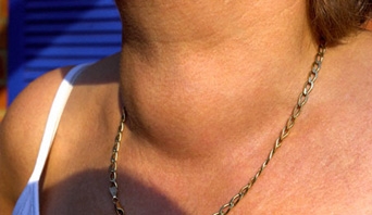
Symptoms of Graves disease


  • Goitre, an enlargement of the thyroid gland, this can be noticed on the neck as a belly like protrusion.

  • Increased metabolic rate, therefore hyperactivity and irritability.
  • Difficulty in sleeping (Insomnia) 
  • Sweating
  • Heat intolerance
  • Graves ophthalmopathy, since the TSH receptors can be found all over our body cells, sometimes the auto-antibodies manage to bind to the TSH receptors on the tissues surrounding the eye. 
  • Weight loss
  • Graves dermopathy


  • Faster heartbeat (Tachycardia) which can lead to congestive heart failure (CHF)
  • Anxiety
  • Hyperreflexia - briefly is the overreaction of the involuntary nervous system to any external stimuli.
  • Hand tremors, an uncontrolled rhythm movement of the hand due to abnormal signals in the brain that miscommunicates with the hands and muscles - hand tremors can also happen due to neurological disorders such as dopamine deficiency like in Parkinson's disease ( you can check out this video ( I do not own it) about Tremor disorders: https://www.youtube.com/watch?v=-Y3kex_8UoY)
References

  • https://www.youtube.com/user/armandohasudungan
  • https://www.youtube.com/user/aimmds
  • https://www.youtube.com/watch?v=SCV_m91mN-Q
  • http://www.yourhormones.info/glands/thyroid-gland/

Next will be - the effect of anabolic steroids on the thyroid hormones and ultimately the metabolism.

Friday, 19 January 2018

Detection of Rutin test in Tea - (Qualitatively)

Detection of Rutin

Qualitatively detected


Introduction

 Rutin is an example of flavanones/flavanoids which should give you the impression of being an antioxidant substance, it is found in a number of fruits and vegetables, it has a wide range of health benefits such as being anticarcinogenic and anti-inflammatory effects.

Chemical structure of Rutin
quercetin-3-rutinoside
Rutin is composed of a disaccharide ( Rhamnose + glucose) glycosylated with a quercetin group ( which is a plant polyphenol compound )

Protein disulfide isomerase is an essential enzyme in protein folding that is synthesized in the endoplasmic reticulum of our cells & platelets and its function is to catalyze the formation and breakage of disulfide bonds - the function of such enzyme is sort of a mixed blessing where performing its job inside the cell is a good thing but when it goes outside the cell it becomes lethal where it can clots proteins which can then accumulate inside the blood vessels, blocking them, ultimately causing Blood clots.

Beside other compounds tested, Rutin was found to be the perfect one to be the inhibitor of Protein disulfide isomerase (PDI) and at the same time it's structure prevent it from entering the cell or otherwise, it would roam around inhibiting any nearby enzyme, so it was said to be anti-thrombotic activity.

Foods rich in Rutin
  • Onion
  • Tea
  • Apples
  • Citrus fruits
  • Buckwheat
Tools
  1. Graduated cylinder (preferably 50ml.)
  2. 2 Droppers 
  3. Ferric chloride solution
  4. 2 test tubes, one kept as a control and the other the test.
  5. Source of flame
Procedures
  1. Weight 1 gram of tea (rutin source)
  2. Add 50ml. of water in a boiler, and leave it till it starts to boil
  3. The onset of boiling put your 1 gram tea.
  4. Leave it to boil for five minutes.
  5. After five minutes of boiling, cool the mixture.
  6. Transfer 1ml. of your tea solution in each of your 2 test tubes.
  7. Add few drops of ferric chloride solution to the test tube and leave the control tube blank.
  8. Record your observation.
Result

  • An olive-green color should be formed as a positive result for the detection of Rutin in tea.

A positive result can be seen in the upper (test) tube comparing it to the control tube

References

  • Food chemistry 15306
  • https://news.harvard.edu/gazette/story/2012/05/flavonoid-compound-can-prevent-blood-clots/
  • https://harvardmagazine.com/2012/09/curbing-clots
  • https://www.healthline.com/health/potential-benefits-of-rutin#diet
  • https://www.medicinenet.com/rutin/supplements-vitamins.htm
  • http://www.phytochemicals.info/phytochemicals/rutin.php
  • https://www.ncbi.nlm.nih.gov/pmc/articles/PMC3739031/

Thanks a lot for reading, I hope you find it informative
#WeliveWelearn


Thursday, 13 July 2017

Ed and Rhizopus stolonifer, an agony tale

Mycology #2

Rhizopus stolonifer (Bread mold)

under general environment

Rhizopus stolonifer has grown on a piece
of bread - the black spots indicate the
formation of sporangium (spore sac)
The fungus was grown under general environment, a piece of bread was cut, put in a small box containing some water, which accelerated the growth of Rhizopus stolonifer or in other words the black mold which usually attacks bread. 

It is considered to be a mesophilic fungus since it rapidly grows between temperatures of 15*C - 30*C.

It's classification, Division: Amastigomycota 
                               Sub-division: Zygomycotina
                                 Class: Zygomycetes

And this class by which this fungus belongs to are characterized by
  1. Terrestrial.
  2. Doesn't form any type of swarm spores ( they are spores that have mobility function (motile) which has a flagellum used for locomotion)
  3. Spores are dispersed either by air or by soil.
  4. Each fungus consists of mycelium ( somatic structure of the body) which form filamentous hyphae  have large proportion of chitin)  - Mycelium is a network of hyphae
  5. The cell wall is formed of chitin
  6. Saprophytic ( little parasitism on plants and animals )
  7. Reproduce asexually by sporangiospores which are non-motile formed within sporangium (sac) reproduce by conidia ( single or chain cells present on the top of conidiophores)
  8. Reproduce sexually by Zygospore due to 2 sex organs similar in shape, size, and aren't differentiated from each other but physiologically different.
The following pictures discuss the structure of the fungus under microscope ( made by me) 
Sporangiophore and sporangium
Structure of Rhizopus stolonifer under microscope


Life cycle of Rhizopus stolonifer
Source: http://bioweb.uwlax.edu/bio203/2011/olbrantz_chri/reproduction.htm

Source: Microbiology course no. 07201

Latest topics
* https://allaboutbiologyworld.blogspot.com/2017/07/detection-of-catalase-in-potatoes.html
* https://allaboutbiologyworld.blogspot.com/2017/07/cob-clot-on-boiling-test.html
* https://allaboutbiologyworld.blogspot.com/2017/06/cell-division-mitosis-meiosis-comparison.html

I hope you find this informative and if you do please follow my blog if you still hadn't and share it with your science buds.

Penicillium digitatum topic - https://allaboutbiologyworld.blogspot.com/2017/04/todd-has-been-sick-short-tale.html

Thanks!

Tuesday, 11 July 2017

Detection of Catalase in potatoes


Detection of Catalase


Catalase  + H2O2  ----------------> water + oxygen


Catalase enzyme is usually found in every oxygen-dependent organism's cells, which catalyzes the decomposition of hydrogen peroxide into water and oxygen, transforming the lethal hydrogen peroxide which is produced normally from the metabolism carried out by our cells into non-lethal compounds ( water & oxygen)

Catalase not only decompose hydrogen peroxide, But compounds like hydrogen peroxides belongs to a category called ''Reactive oxygen species'' or ROS, they are oxidizing species of high energy, containing oxygen, unstable, easily react with other chemical compounds like our DNA damaging it, They are often radicals meaning they contain unpaired electrons such as H2O2, OH ( hydroxyl radical ), and superoxides.


They are produced by cleavage of water with ionizing radiation (ultraviolet and higher energies). They are also normally produced in cellular metabolism and immediately destroyed by other enzymes, but many pollutants, such as tobacco smoke, cause increase in their production - when more is produced than can be removed, they have time to cause DNA damage.

The bubbles produced are the oxygen molecules due to
decomposition of hydrogen peroxide
Tools
  • Hydrogen peroxide bottle
  • Test tube
  • Dropper
  • Small pieces of cut potato so they can fit inside the test tube
Procedures
  1. Cut very small pieces the size of the test tube's diameter you are going to perform the test in so they can fit inside.
  2. Use your dropper to take a few milliliters of hydrogen peroxide like 3 to 5 ml. 
  3. Put the small pieces of potato inside the test tube then pour the content of the dropper inside and watch the reaction performed.
Potatoes are living plant tissue even when harvested so they do respire oxygen normally after being harvested so they have Catalase which when hydrogen peroxide is poured onto it, it will start to bind to the hydrogen peroxide forming a complex that will decompose the hydrogen peroxide to water and oxygen making it non-lethal.

I hope you gentlemen and ladies find this informative and if you do please do share it with your science buds and follow my blog for new topics.

I will upload a complete video explaining all the steps in case you find something difficult or how you going to perform this test at home - so the video COMING SOON...

Recent topic - https://allaboutbiologyworld.blogspot.com/2017/06/cell-division-mitosis-meiosis-comparison.html
https://allaboutbiologyworld.blogspot.com/2017/07/cob-clot-on-boiling-test.html

#Biology #Catalase
#WeliveWelearn

Monday, 10 July 2017

C.O.B - Clot On Boiling Test

Dairy Science

Clot . On . Boiling Test

Determination of Milk Acidity

Introduction
The normal pH of a natural milk produced ranges from 6.6 to 6.8 and of an acidity 0.13 to 0.18, this acidity is called the Natural Acidity due to some constituents which make up this acidity in milk such as Casein ( composed of α-casein, β-casein, and γ-casein), Whey protein ( composed of Lactoglobulin and Lactoalbumin), Salts ( e.g., Phosphates, Citrates, Nitrogen, Potassium, Zinc, Boron, etc...) and CO2

Leaving milk under normal temperature such as room temperature, this allows for the growth of bacteria present in the milk especially the Lactic acid bacterial strains which converts the Lactose ( milk sugar) to Lactic acid by fermentation decreasing the pH and increasing the milk acidity, and this acidity is called Developed Acidity; Because it came/rose or developed from an organism which is in our case the Lactic acid bacteria.

There are qualitative and quantitative methods of acidity determination for milk bulks in factories/plants and usually we measure the acidity by measuring lactic acid percentage, and in this case we call it '' Total Titratable Acidity ''.

Why do we need to determine acidity in milk?
Because it is important for the manufacture of some dairy products, quality of milk, and the milk stability during some technological treatments.

It is a qualitative method of acidity determination in milk, we conclude from the test name is that we are counting on the formation or the appearance of Clots in the milk sample, so the milk will be clotted if acidity is 0.25% and more

Tools needed ( Home )
  • Pipette
  • Beaker ( will be used as a hot water bath)
  • Test tube
  • Milk sample
  • Flame source

Procedures
  1. Put 5 ml. of milk in clean test tube.
  2. Put the test tube in boiling water bath until boiling of the sample.
  3. Notice the presence of clots, if present it will be positive result, If not, then it will be a negative result.


+ve result - By me 
The formation of clots indicates the high acidity of the milk, and perhaps you could ask how I received an acidic milk sample:
 Basically, I took like an amount of 200 ml., and a tablespoon of yoghurt ( containing a Lactic acid bacteria Bifidobacterium sp., and Lactobacillus sp.), and left them for about a day in room temperature until a curd was formed. Yet the whole sample wasn't curd, Once a curd was formed I managed to take 5 ml. and perform the test.

I hope you gentlemen and ladies find this informative and if you do please do share it with your science buds and follow my blog for new topics.

I will upload a complete video explaining all the steps in case you find something difficult or how you going to perform this test at home - so the video COMING SOON...

Next one in our dairy science episodes laboratory work experiments will be the quantitative method of milk acidity determination..., the scientific method. STAY TUNED

Recent topic - https://allaboutbiologyworld.blogspot.com/2017/06/cell-division-mitosis-meiosis-comparison.html

#Biology #DairyScience
#WeliveWelearn


Wednesday, 28 June 2017

Cell division - Mitosis & Meiosis comparison

Mitosis Vs. Meiosis


Points of comparison
Mitosis
Meiosis
Stages
Lasts in 4 stages
Lasts in 8 stages
Separation of chromosomes
Identical chromatids separate as independent chromosomes
Homologous chromosomes ( each with 2 chromatids ) separate
Pairing of chromosomes
No pairing occurs
Pairing occurs to form tetrads
Number of the resulting cells
2 diploid cells (2n each)
4 haploid gametes ( n each)
Occurs in
Somatic cells
Gonads
Crossing over
Does not occur
Occurs
Chiasmata
Does not occur
Observed during Prophase l and Metaphase l
Prophase


Resulting genetic characters
Identical in each cell & as the mother cell
Each gamete has different characters due to crossing over which allows for genetic variation
Karyokinesis
Occur in Interphase
Occur in Interphase l
Cytokinesis
Occur in Telophase
Occur in Telophase l and in Telophase ll
Phases happen
Once
Twice
Prophase
It usually takes a couple of hours, and therefore it is simple
It takes longer time comparing to the one in mitosis up to a day, and therefore it is complicated
Synapsis
Does not occur
Happens in homologous chromosomes during Prophase
Chromosomal arrangement during metaphase
Chromosomes arrange in one row and centromere divides
Chromosomes arrange in 2 rows & centromere does not divide ( Meiosis  I )
Location of occurrence
In plants: growing tips ( apical meristems in shoots and roots) & cambium layer ( meristematic cells) which give rise to xylem and phloem tissues
In animals: all cells except nerve cells
In plants: anthers & ovaries of flowers
In animals: testis & ovaries.
Considered to be       
A building method of the human body especially in the phases of early development (during Zygote stage - Mitosis is so active then it continues to be active after delivery until it becomes regular) replenishing method for worn out tissues or regeneration of new layers such as your skin epidermal layer.
It is also considered as a reproduction method for some organisms ''Asexual reproduction''
Aims for the reduction of the chromosomal number and genetic variation of organisms - it is carried out by higher advanced organisms in life
''Sexual reproduction''
Discovered by
Walther Flemming
Oscar Hertwig ( described by him for the first time) then by Edouard Van Beneden ( described it for the second time)
Diagram


**************The End**************


Notice: All pictures inside this topic doesn't belong to me - copyrights goes to the owner:
http://bio1510.biology.gatech.edu/
http://www.ducksters.com/science/biology/cell_division.php
https://www.pinterest.com/peggleston22/science/
local-brookings.k12.sd.us 

Recent topics
Difference between animal and plant cells 
http://allaboutbiologyworld.blogspot.com/2017/06/difference-between-animal-and-plant.html

Overview of plant cells - part 1 Structure and function
http://allaboutbiologyworld.blogspot.com/2017/06/overview-of-plant-cells-part-1.html

Difference between Eukaryotes and Prokaryotes
http://allaboutbiologyworld.blogspot.com/2017/06/a-comparison-between-prokaryotes-and.html

Tuesday, 27 June 2017

Difference between Animal and Plant cells

Animal cells Vs. Plant cells

Points of comparison
Animal cell
Plant cell
Shape
Irregular due to possessing only cell membrane
Rigid in shape due to cell wall which consist of compounds that give the cell wall its rigidity
Organelles
Has centrioles, and lysosomes
Small vacuoles relative to those found in plant cells
Lack chloroplasts and cell wall
Has chloroplast( organelle containing pigment chlorophyll which gives the plants their green color), cell wall and central large vacuole.
Lack centrioles
Totipotency
Only special types of cells can regenerate or differentiate to specific type of cells '' these cells are stem cells'' (Embryonic stem cells (Pluripotent) & somatic stem cells ( adult stem cells or multipotent stem cells)
Can originate a whole new complete plant from any single cell
Genes
All genes are found in all cells but not all of them are active e.g., Insulin-producing gene is active in the pancreas and inactive in your lung cells
All genes are active since they have the ability to regenerate from one single cells a complete plant ''Totipotency''
Metabolism
Animal cells cannot perform photosynthesis since they lack chloroplasts
Plants have one specific different process in their life cycle that no other organisms has it ''Beside some algae and bacterial strains''
Which is the photosynthesis due to chloroplasts
During mitotic cell division
In Prophase, a centrosome ( has 2 pairs of centrioles) is found near the nucleus. Each centriole moves to one end of the cell forming spindle fibers.
In Telophase, As the nucleus is now completely divided, the cytoplasm start to divide, a constriction appears & continues in the middle of the cell dividing it into 2 cells.
In Prophase, No centrioles are found. Spindle fibers are formed directly from the cytoplasm
In Telophase, No constriction, a middle lamella is formed to separate the formed cells. The middle lamella is in the form of a chain of fine vesicles followed by the precipitation of pectic compounds and then cellulose is followed forming the cell wall separating plant cells.
Nucleus
It is usually in the middle of the cell
It is usually not centered due to the relatively large size of the central vacuole taking up much space.
Starch granules
Absent - excess glucose is usually connected together in chains forming chains of glycogen which are stored in skeletal muscles and liver for later use
Present
Diagram


Recent topics

Overview of plant cells - part 1 structure and function -http://allaboutbiologyworld.blogspot.com/2017/06/overview-of-plant-cells-part-1.html


Difference between Eukaryotes and Prokaryotes http://allaboutbiologyworld.blogspot.com/2017/06/a-comparison-between-prokaryotes-and.html


Animal cells - structure of the organelles and their functions 
http://allaboutbiologyworld.blogspot.com/2017/06/our-cell-1-structure-function-division.html 

Biology gallery #2 - http://allaboutbiologyworld.blogspot.com/2017/06/biology-gallery-2.html